Lunit has highlighted in this recent and first retrospective reader study its potential to enhance breast cancer screening pathways with AI. The study involved the interpretation of digital breast tomosynthesis (DBT) scans from 258 women by eight general radiologists and seven breast specialists and it found that AI assistance allowed general radiologists to achieve diagnostic performances comparable to those of breast specialists. With Lunit INSIGHT DBT, the overall AUC for radiologists improved from 0.90 to 0.92 and the reading time decreased from 54.41 seconds to 48.52 seconds. AI support increased sensitivity, particularly in fatty breasts, and specificity in dense breasts. Additionally, the study revealed that with AI improved radiologists’ performance in detecting and diagnosing soft tissue lesions compared to calcifications alone, emphasizing its utility in identifying soft tissue lesions on DBT. Furthermore, interreader agreement improved when using AI. These findings suggest a promising role for AI in DBT diagnostic precision and streamlining workflow in breast cancer detection.
Read full study
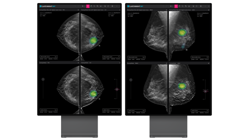
Impact of AI for Digital Breast Tomosynthesis on Breast Cancer Detection and Interpretation Time
Radiology: Artificial Intelligence, 2024
Abstract
Objective: To develop an artificial intelligence (AI) for diagnosis of breast cancer in digital breast tomosynthesis (DBT) and investigate whether it could improve diagnostic accuracy and reduce reading time of radiologists.
Materials and methods: A deep learning AI algorithm was developed and validated for DBT with retrospectively collected examinations (January 2010 to December 2021) from 14 institutions in the United States and South Korea. A multicenter, reader study was performed to compare the performance of 15 radiologists (7 breast specialists, 8 general radiologists) in interpreting DBT examinations from 258 women (mean, 56 years ± 13.41 [SD]), including 65 cancer cases, with and without the use of AI. Area under the receiver operating characteristic curve (AUC), sensitivity, specificity, and reading time were evaluated.
Results: The AUC for standalone AI performance was 0.93 (95% CI: 0.92,0.94). With AI, radiologists’ AUC improved from 0.90 (0.86, 0.93) to 0.92 (0.88, 0.96; P = .003) in the reader study. AI showed higher specificity (89.64% (85.34, 93.94)) than radiologists (77.34% (75.82, 78.87; P < .001)). When reading with AI, radiologists’ sensitivity increased from 85.44% (83.22, 87.65) to 87.69% (85.63, 89.75; P = .04), with no evidence of a difference in specificity. Reading time decreased from 54.41 seconds (52.56, 56.27) without AI to 48.52 seconds (46.79, 50.25) with AI (P < .001). Interreader agreement measured by Fleiss kappa increased from 0.59 to 0.62, respectively.
Conclusion: The AI model showed better diagnostic accuracy than radiologists in breast cancer detection and reduced reading times. The concurrent use of AI in DBT interpretation could improve both accuracy and efficiency.