The AI software Boneview Trauma by Gleamer was evaluated for its effectiveness in enhancing fracture detection. The work involved 101 patients who underwent radiographic examinations following trauma, with the AI being tested both as a standalone tool and as an aid for a junior and a senior radiologist. The findings demonstrated that AI significantly improved the fracture detection capabilities of both junior and senior radiologists, particularly for common fracture types. However, for subtle fractures, the software's performance did not surpass that of experienced radiologists. This highlights the potential of AI as a supportive tool, especially beneficial for less experienced radiologists, and suggests that while AI can enhance diagnostic accuracy, it may not replace the expertise of expert radiologists in complex cases (yet).
Read full study
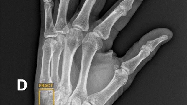
Radiographic Detection of Post-Traumatic Bone Fractures: Contribution of Artificial Intelligence Software to the Analysis of Senior and Junior Radiologists
Journal of the Belgian Society of Radiology, 2024
Abstract
Objective: The aims of this study were: (a) to evaluate the performance of an artificial intelligence (AI) software package (Boneview Trauma, Gleamer) for the detection of post-traumatic bone fractures in radiography as a standalone; (b) used by two radiologists (osteoarticular senior and junior); and (c) to determine to whom AI would be most helpful.
Materials and methods: Within 14 days of a trauma, 101 consecutive patients underwent radiographic examination of the upper or lower limbs. The definite diagnosis for identifying fractures was: (a) radio-clinical consensus between the radiologist on-call who analyzed the images and the orthopedist (Group 1); (b) Cone Beam computed tomography (CBCT) exploration of the area of interest, in case of doubts or absence of consensus (Group 2). Independently of this diagnosis for both groups, the radiographic images were separately analyzed by two radiologists (osteoarticular senior: SR; junior: JR) prior without, and thereafter with the results of AI.
Results: AI performed better than the radiologists in detecting common fractures (Group 1), but not subtle fractures (Group 2). In association with AI, both radiologists increased their overall performances in both groups, whereas this increase was significantly higher for the JR (p < 0.05).
Conclusion: AI is reliable for common radiographic fracture identification and is a useful learning tool for radiologists in training. However, the software's overall performance does not exceed that of an osteoarticular senior radiologist, particularly in case of subtle lesions.