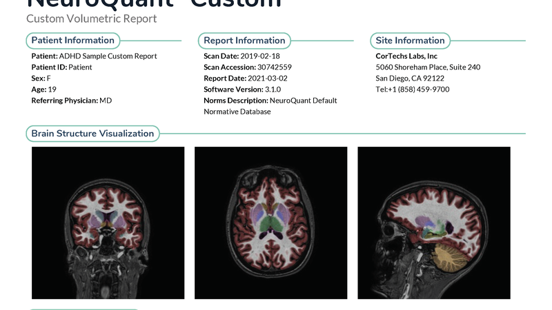
NeuroQuant (NQ) automated volumetry capabilities were evaluated in this study, compared against traditional visual rating scales for diagnosing dementia. Researchers at Oslo University Hospital’s Memory Clinic analyzed brain magnetic resonance examinations from 86 patients, assessing both NQ and visual scales that measured medial temporal lobe atrophy, global cortical atrophy, and posterior atrophy. ROC analyses were performed to validate the performance of various MRI classifiers - including three visual measures and five NQ volumetrics - in distinguishing dementia from non-dementia. The analyses showed that conjoining all NQ volumes resulted in an AUC of 0.77. Furthermore, NQ volumetrics generally matched or exceeded the diagnostic accuracy of visual ratings, with NQ’s hippocampal volume measurements were particularly effective, displaying higher diagnostic accuracy than visual assessments of the same regions. Integrating both NQ volumetrics and visual ratings improved diagnostic predictions for posterior brain regions, suggesting that combining automated and traditional methods could enhance diagnostic precision in clinical settings.
Read full study
Regional MRI volumetry using NeuroQuant versus visual rating scales in patients with cognitive impairment and dementia
Brain and Behavior, 2024
Abstract
Objective: The aims were to compare the novel regional brain volumetric measures derived by the automatic software NeuroQuant (NQ) with clinically used visual rating scales of medial temporal lobe atrophy (MTA), global cortical atrophy-frontal (GCA-f), and posterior atrophy (PA) brain regions, assessing their diagnostic validity, and to explore if combining automatic and visual methods would increase diagnostic prediction accuracy.
Material and methods: Brain magnetic resonance imaging (MRI) examinations from 86 patients with subjective and mild cognitive impairment (i.e., non-dementia, n = 41) and dementia (n = 45) from the Memory Clinic at Oslo University Hospital were assessed using NQ volumetry and with visual rating scales. Correlations, receiver operating characteristic analyses calculating area under the curves (AUCs) for diagnostic accuracy, and logistic regression analyses were performed.
Results: The correlations between NQ volumetrics and visual ratings of corresponding regions were generally high between NQ hippocampi/temporal volumes and MTA (r = -0.72/-0.65) and between NQ frontal volume and GCA-f (r = -0.62) but lower between NQ parietal/occipital volumes and PA (r = -0.49/-0.37). AUCs of each region, separating non-dementia from dementia, were generally comparable between the two methods, except that NQ hippocampi volume did substantially better than visual MTA (AUC = 0.80 vs. 0.69). Combining both MRI methods increased only the explained variance of the diagnostic prediction substantially regarding the posterior brain region.
Conclusions: The findings of this study encourage the use of regional automatic volumetry in locations lacking neuroradiologists with experience in the rating of atrophy typical of neurodegenerative diseases, and in primary care settings.